Infections in prostheses, for amputees, and in joint replacements, are common, often found too late, and can necessitate additional surgeries or worse complications. Current detection methods include MRI, CT, and X-ray.
Ken Loh and UCSD colleagues have developed an infection detecting prosthesis coating + imaging technique that could be used at home or in a doctors office. Prostheses are continuously monitored and quantitative diagnostic data about the extent and location of an infection is provided.
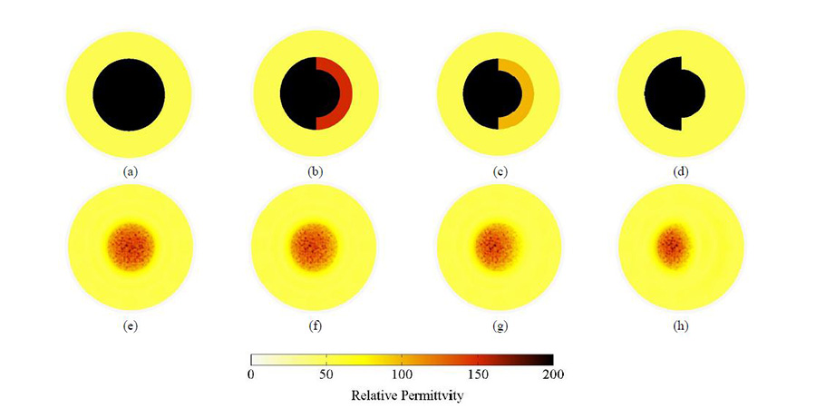
Enhanced ECT was used to measure the human tissue and prosthesis’ electrical properties using electrical fields. An algorithm processed the data to allow physicians to reconstruct a predetermined area’s electrical properties, to reveal tissue, bone, and prosthesis health. Infection caused changes in the field, detected by ECT.
A thin-film sensor was sprayed onto a prosthesis to improve infection detection. The film is made of a conductive polymer matrix that is sensitive to pH, and carbon nanotubes, embedded in the matrix, to increase the material’s ability to conduct electricity more sensitively, regardless of the pH level. Infections caused by different microorganisms often change the local pH in human cells and affect their ability to conduct electricity.
ApplySci’s 6th Digital Health + NeuroTech Silicon Valley – February 7-8 2017 @ Stanford | Featuring: Vinod Khosla – Tom Insel – Zhenan Bao – Phillip Alvelda – Nathan Intrator – John Rogers – Roozbeh Ghaffari –Tarun Wadhwa – Eythor Bender – Unity Stoakes – Mounir Zok – Sky Christopherson – Marcus Weldon – Krishna Shenoy – Karl Deisseroth – Shahin Farshchi – Casper de Clercq – Mary Lou Jepsen – Vivek Wadhwa – Dirk Schapeler – Miguel Nicolelis